This job description has been automatically translated. Please note that automatic translation is not 100% accurate, so there may be minor translation errors in the text.
| Instructions for the position " Laboratory assistant for pathological research", presented on the website, meets the requirements of the document - "DIRECTORY qualification characteristics workers' professions. Issue 78. Healthcare. (As amended in accordance with orders of the Ministry of Health No. 131-O dated June 18, 2003, No. 277 dated May 25, 2007, No. 153 dated March 21, 2011, No. 121 dated February 14, 2012)", which was approved by order of the Ministry of Health of Ukraine on March 29, 2002 N 117. Agreed by the Ministry of Labor and social policy Ukraine. The document status is "valid". |
|
Preface to the job description
0.1. The document comes into force from the moment of approval.
0.2. Document developer: _ _ _ _ _ _ _ _ _ _ _ _ _ _ _ _ _ _ _ _ _ _ _.
0.3. The document has been approved: _ _ _ _ _ _ _ _ _ _ _ _ _ _ _ _ _ _ _ _ _ _ _ _.
0.4. Periodic verification of this document is carried out at intervals not exceeding 3 years.
1. General provisions
1.1. The position "Pathological Research Laboratory Assistant" belongs to the "Specialists" category.
1.2. Qualification requirements- incomplete higher education(junior specialist) or basic higher education (bachelor) in the field of preparation "Medicine", specialty "Laboratory diagnostics". Specialization in Pathological Anatomy. No work experience requirements.
1.3. Knows and applies in practice:
- current legislation on health protection and regulatory documents regulating the activities of health care institutions;
- organizing the work of the pathology laboratory;
- rights, duties and responsibilities of a laboratory assistant for pathological research;
- basics of histology and cytology;
- rules for collecting material, preparing tissue sections on a microtome, storing, fixing;
- fixing media, dehydration agents;
- principles of preparation of paint reagents, absolute alcohol, its dilution;
- techniques for embedding in paraffin, painting frozen and paraffin sections, collagen and elastic fibers, nervous tissue;
- histochemical methods for studying nucleic acids, enzymes, etc.;
- rules for the operation of laboratory equipment and labor protection in the laboratory;
- principles of first and urgent care medical care;
- basic medicines, dosages and methods of their introduction into the body in emergency conditions;
- principles of anti-epidemic regime in the laboratory;
- rules for preparing medical documentation.
1.4. A pathological research laboratory assistant is appointed and dismissed by order of the organization (enterprise/institution).
1.5. The pathology laboratory assistant reports directly to _ _ _ _ _ _ _ _ _ _ .
1.6. The pathological research laboratory assistant supervises the work of _ _ _ _ _ _ _ _ _ _ .
1.7. During absence, the pathological research laboratory assistant is replaced by a person appointed in accordance with the established procedure, who acquires the appropriate rights and is responsible for the proper performance of the duties assigned to him.
2. Characteristics of work, tasks and job responsibilities
2.1. Guided by the current legislation of Ukraine on health care and regulations that determine the activities of healthcare institutions and the organization of work of pathological laboratories.
2.2. Selects material for histological studies, fixes it, stores it, and fills the material for electron microscopic studies.
2.3. Prepares tissue sections, masters the technique of embedding in paraffin, glues paraffin sections onto a glass slide, and carries out preliminary preparation of paraffin.
2.4. Prepares paints, reagents, utensils, and equipment.
2.5. Knows how to work on microtomes, knows how to prepare microtome knives.
2.6. Adheres to the principles of medical deontology.
2.7. Maintains medical records.
2.8. Constantly improves his professional level.
2.9. Knows, understands and applies current regulations relating to his activities.
2.10. Knows and complies with the requirements of regulations on labor protection and environmental protection, complies with the norms, methods and techniques for the safe performance of work.
3. Rights
3.1. The pathology technician has the authority to take action to prevent and correct any irregularities or inconsistencies.
3.2. A pathological research laboratory technician has the right to receive all provided for by law social guarantees.
3.3. A pathological research laboratory technician has the right to demand assistance in the performance of his duties and the exercise of his rights.
3.4. A pathological research laboratory technician has the right to demand the creation of organizational and technical conditions necessary for the performance of official duties and the provision necessary equipment and inventory.
3.5. A pathological research laboratory technician has the right to familiarize himself with draft documents relating to his activities.
3.6. A pathological research laboratory technician has the right to request and receive documents, materials and information necessary to perform his job duties and management orders.
3.7. A pathological research laboratory technician has the right to improve his professional qualifications.
3.8. A pathological research laboratory technician has the right to report all violations and inconsistencies identified in the course of his activities and make proposals for their elimination.
3.9. A pathological research laboratory technician has the right to familiarize himself with documents defining the rights and responsibilities of his position, and criteria for assessing the quality of performance of official duties.
4. Responsibility
4.1. The pathological research laboratory assistant is responsible for failure to fulfill or untimely fulfillment of the requirements of this job description obligations and (or) non-use of granted rights.
4.2. The pathological research laboratory assistant is responsible for failure to comply with internal rules. labor regulations, occupational health, safety, industrial sanitation and fire protection.
4.3. The pathological research laboratory assistant is responsible for disclosing information about the organization (enterprise/institution) that is a trade secret.
4.4. The pathological research laboratory assistant is responsible for non-fulfillment or improper fulfillment of the requirements of internal regulatory documents of the organization (enterprise/institution) and legal orders of management.
4.5. The pathological research laboratory assistant is responsible for offenses committed in the course of his activities, within the limits established by the current administrative, criminal and civil legislation.
4.6. The pathological research laboratory technician is responsible for causing material damage to the organization (enterprise/institution) within the limits established by the current administrative, criminal and civil legislation.
4.7. The pathological research laboratory assistant is responsible for the unlawful use of the granted official powers, as well as their use for personal purposes.
more than 6 months without presenting requirements for work experience.
Taking this into account, the administration of the State Budgetary Institution of Healthcare of the Moscow Region “Bureau of Forensic Medicine”, on the basis of Order of the Ministry of Health and Social Development of the Russian Federation No. 541 of July 23, 2010 “On approval of the Unified Qualification Directory of Positions of Managers, Specialists and Employees”, decided to train all medical registrars who do not have a secondary medical education, with a degree in medical registrar. Specialists from Platon Consulting Group LLC, who have a license for this type activities. 80 people received on-the-job training.
Medical registrars studied the basics of labor and labor protection legislation of the Russian Federation; internal labor regulations of the Bureau of Emergency Management; fundamentals of the legislation of the Russian Federation on protecting the health of citizens; requirements professional standard and job responsibilities of a medical registrar. During the training process, medical registrars mastered the basics professional activity, including the procedure for registering and receiving cadaveric material in accordance with Order of the Ministry of Health of the Russian Federation No. 346 of May 12, 2010 “On approval of the procedure for organizing and conducting forensic medical examinations in state forensic institutions of the Russian Federation”; acquired first aid skills; mastered working with computer information systems. Classes were conducted in the form of distance lectures, tests, independent studies followed by passing qualifying exams.
Quality of work of the State budgetary institution Healthcare of the Moscow Region "Bureau of Forensic Medicine" depends on the quality of administration at all levels, from the head of the bureau to medical workers of the second and third echelons, including medical registrars. Having received appropriate training, medical registrars can perform their functional responsibilities at the proper level.
LABORATORY HISTOLOGIST: YESTERDAY AND TODAY
L. V. Danchenko
Bureau of Forensic Medicine of the Moscow Region (head - Doctor of Medical Sciences, Prof. A. A. Klevno)
Abstract: The report is devoted to the peculiarities of the work of a laboratory assistant in the histological department
at the present stage of development of histology as a medical discipline.
Key words: histology, laboratory assistant, laboratory, histological specimen, diagnostics
INTRODUCTION
Histology is the science of studying tissues and organs using a microscope. This medical discipline is an integral part of pathological anatomy and forensic medicine. Histology as a science received its active development in the 18th century, and in the 19th century it became an integral part of the diagnosis of various pathological processes. It was during this period that the need arose for the emergence of such a specialty as a laboratory assistant. The main task of the histologist laboratory assistant is to produce
preparation of histological preparations from pieces of organs and tissues, which the doctor subsequently examines under a microscope. Since the emergence of histology as a discipline to this day, the basic principle of preparing the drug remains the same.
Processing tissues taken for histological examination is a very labor-intensive, complex and lengthy process. On average, the preparation time for the drug can take 5-7 days. This is due to the fact that this process is still carried out manually. Various types of fabrics are used for processing chemicals, many of them are toxic. Under these conditions, various errors in technological process. All this affects the quality of the histological specimen and, as a consequence, the quality of the histological examination itself, which is carried out by the doctor. Therefore, the laboratory assistant is required to have special theoretical knowledge, strict adherence to all stages and time parameters of tissue processing, as well as possession of special practical skills. At the same time, the laboratory histologist must not only be professionally trained, but also have attention, perseverance, patience and responsibility.
In the last decade, there has been active development of the histological research method. This is due to its widespread use in intravital diagnostics. various diseases in pathological anatomy. IN forensic medicine the increase in the number of histological studies is associated with a change in the structure of mortality (the predominance of non-violent), the complexity of the material entering the histological department and the range of issues to be resolved. To expand the capabilities of the histological research method, the laboratory histologist must have the skills to conduct various types colors The recommended list of stains, which allows solving more diagnostic problems, includes more than 20 items. All this increases the volume of histological examination due to an increase in the number of preparations. However, the number of preparations and additional staining techniques are not taken into account when determining the laboratory assistant’s workload. In addition, the number of available rates often does not correspond to the volume of work performed in the laboratories. All this, unfortunately, does not stimulate the introduction of additional new stains and methods of histological examination (morphometry, polarization microscopy, etc.).
In order to facilitate the work of the laboratory assistant and due to the need for more short terms and carry out quality large number histological studies, over the past 10-15 years, semi-automatic and automatic devices have been actively developed that can be used at every stage of material processing. The transition from a manual to an automatic method of tissue processing allows you to maximize the quality and reduce the time of the process of fixing, wiring, pouring and staining of preparations. These devices can be used around the clock and have a delayed start. This reduces the production time for histological preparations from 7 to 3-4 days, even in cases where a large number of research objects are received.
In connection with the introduction of new technologies in the drug production process, new requirements are placed on the histology department laboratory assistant. These are additional specialized knowledge to work with complex technology and computer, programming skills. General knowledge of the structure of tissues and organs, basic general pathological processes is necessary. This is due to the introduction into practice of using
control by laboratory assistants themselves of the quality of manufactured histological preparations under a microscope.
Thus, I would like to note that at the present stage, the capabilities of the histological research method are not fully used everywhere. This is associated with many difficulties, and primarily with financing opportunities. To introduce new technologies and increase the number of studies conducted, high-tech equipment and consumables are required, which requires large financial investments that not every medical institution can afford.
Submitting your good work to the knowledge base is easy. Use the form below
Students, graduate students, young scientists who use the knowledge base in their studies and work will be very grateful to you.
Posted on http://www.allbest.ru/
Introduction
Chapter 1. Analysis of some factors determining the development and effectiveness of the work of a histologist laboratory assistant
2.4 Planning work taking into account the technical structure of the laboratory
2.5 Selection of technologies and equipment for the histology laboratory assistant’s workplace
2.6 Integrated technological solutions in the work of a histologist in the laboratory
Conclusion
References
Glossary
Introduction
laboratory assistant histologist biopsy risk
Histology (the term is formed by combining the Greek words “histos” - “tissue” and “-logia” - “science”) is the study of the anatomy of cells and tissues of representatives of the plant and animal world on a microscopic scale. Typically, it is performed by analyzing tissues and cells using sectioning and staining, followed by examination under a light or electron microscope. Histological studies can be carried out using tissue culture, where living cells can be isolated and maintained in a proper environment outside the body in various research activities.
Histology, or more precisely histopathology, is a section of the scientific and applied discipline “pathological anatomy” that directly studies pathological changes in tissue to determine the patient’s condition.
The purpose of this work is to study the work of a histologist laboratory assistant, analyze the work and develop individual proposals, create a new technique, technology; opportunities for labor optimization.
Relevance of the study. The development of technology in medicine leads to the expansion of the capabilities of the histology laboratory.
In modern world medicine, the main activity of the laboratory of histology and cytology is aimed at intravital morphological diagnosis of pathological conditions of various organs and systems - current and forward planning at work. This requires equipping such a laboratory with the latest automatic equipment. Here, histological and cytological examination of surgical and biopsy material is performed. Nowadays, doctors need to provide the patient with the necessary and timely assistance in treatment. The time frame for obtaining research results is being accelerated (within 1 working day, and for surgical material within 1-2 working days), immunohistochemical methods for studying infectious pathology are being introduced. For example, it is currently possible to diagnose herpes simplex virus types 1 and 2, cytomegalovirus, Epstein-Barr virus, human papillomavirus (oncogenic strains), parvovirus B19, pneumocystiscarinii, toxoplasma (Toxoplasmagondii), adenoviral infection in biopsy, surgical and cytological material. Despite actions aimed at occupational safety, laboratory assistants who come into contact with histological material run the risk of contracting infections.
Such a laboratory should have a comfortable and modern system archiving materials received from patients, intended for long-term storage and quickly search for diagnostic drugs if necessary.
The laboratory must be staffed by qualified pathologists. Histology laboratory assistants actively help them.
The role of a histologist laboratory assistant in such working conditions currently requires not only theoretical literacy, but also a certain optimization and modernization of work, which will have a positive impact on the work of the laboratory as a whole.
The object of the study is to consider the algorithm of the work of a histology laboratory assistant, the presence of risk factors in the work of a histology laboratory assistant.
The subject of the study is the content, forms and types of activities of secondary medical personnel laboratories.
Research objectives:
study the regulatory framework regulating the work of histological laboratories.
study methods of histological examination and equipment of histological laboratories.
Scientific novelty of the research.
Despite the relevance of the problem, in this work we will try to analyze in detail the risk factors in the work of a histology laboratory assistant.
Practical significance of the study.
Histological diagnosis is developing and becoming more and more in demand, but the analysis process is not automated.
Although the appearance disposable tableware and technical equipment has reduced the risk of infection of medical personnel; it is not possible to avoid it.
Chemical reagents are also not safe; among them there are toxic substances and fire hazards. The transition to modern analyzers has simplified the work of laboratories, however, the lack of modern equipment and interruptions in supply consumables for them it is not possible to completely abandon the old methods.
Microscopy increases eye strain, and if the lighting is insufficient, the risk of decreased vision increases. Insufficient equipment in the premises also adversely affects the health of laboratory staff.
Identification of risk factors and determination of methods for preventing diseases associated with the working conditions of a histology laboratory technician is an urgent problem of modern occupational medicine.
At the moment, there are absolutely not enough pathologists and histology laboratory technicians to fill vacancies in government health care structures.
Hypothesis:
Rational organization of the work of a histologist laboratory assistant, based on approved standards and work procedures, application and mastery of work using latest technologies improves the quality of work, this is directly facilitated by saving time, saving labor costs, improving labor ergonomics, and optimizing the work of a histologist laboratory assistant.
Chapter 1. Analysis of some factors determining the development and effectiveness of the work of a histologist laboratory assistant (literature review)
The main task of practical healthcare is intravital morphological diagnosis. This is the main and important scientific and practical way to solve the diagnostic problems of many specialists. Intravital morphological biopsy functionally and organizationally requires solving many problems.
It must solve many public health issues. The organization must be correct in structure and meet the needs of the clinic at optimal costs.
Pathoanatomical department (synonym prosectura, from Latin prosecare - to dissect) is part of a medical (research) institution in which macro- and microscopic, and in the presence of special rooms, bacteriological, chemical and x-ray examinations of corpses, morphological studies are carried out surgical and biopsy materials.
Organization and equipment of the pathohistology laboratory
According to Order No. 468-64, “rules for the arrangement and operation of the premises of pathology departments and morgues (pathohistological and forensic histological laboratories) of treatment, preventive and forensic medical institutions, institutions and educational institutions(approved by the Ministry of Health)"
The head of the pathology department and (or) histology laboratory must develop based on these rules. The area where the department will be located should be located away from the medical buildings, separated by a forest protection zone - a park or garden with a width of more than 15 m.
The site must be equipped with access roads, a separate entrance, required, as a rule, only for the use of the pathology department and histological laboratory. In some cases, it can only be combined with the entrance to the economic zone.
The pathoanatomical and forensic medical buildings and the entrances to them should not be visible from the windows of the patient premises and from the garden for patients, isolated from adjacent residential buildings.
The department and the morgue cannot be located in the same building with auxiliary services of institutions or treatment rooms and must have separate premises.
The department's premises consist of a sectional room where autopsies are performed, laboratory premises in which material for sections and biopsies is prepared and processed; offices of the director and doctors and from a number of utility rooms: the pre-section room, rooms for storing and issuing corpses, a waiting room for relatives of the deceased, an inventory room, a locker room for employees with individual cabinets, etc. For storing and issuing corpses of those who died from infectious diseases allocate isolated rooms with separate access to the street.
The total area of the premises of the pathology department in hospitals with up to 100 beds is assumed to be 44 square meters. m, and in hospitals with a number of beds of 100 or more based on 1 bed. In hospitals with 100 beds, 1.08 sq. m The area and set of morgue premises are determined based on the population of these cities
The pathology department should be a facility for storing corpses at low temperatures. When storing corpses in a basement or semi-basement, an elevator is required to lift them into the section and return them. The dissection room must satisfy three main conditions: free and well-lit places for the autopsy, comfortable and sufficiently sized places for doctors and students present during the autopsy, convenient access to the dissection tables with stretchers and gurneys. The sectional room is usually located on the first floor. Its area depends on the number of sectional tables (at least 15 m2 per table in small hospitals and 25 m2 in clinical ones). The floor and walls are tiled. The sectional tables and two large rectangular sinks should have cold and hot water. In large medical institutions there may be several sections - large, small and for the corpses of those who died from infectious diseases.
The height of the main rooms in the pathology department and in the morgue should be 3 m.
The layout of the building of the pathology department and the morgue must comply with the following requirements. Separated by a vestibule or corridor from the histological laboratory, rooms associated with the transportation of corpses within the building. Premises for autopsy, processing and storage of unfixed sectional and biopsy material, separate rooms for doctors and service personnel, a museum, a shower and sanitary unit, as well as other premises.
All rooms in the building for the pathology department and histology laboratory must be dry.
A shower room should be provided for the medical staff of the department and morgue; in medical institutions with a number of beds of 400 or more, a sanitary passage is provided.
The walls and partitions are constructed from waterproof inorganic materials.
The walls of the offices should be painted with oil paint up to half the height, and the walls of the sectional, pre-sectional, room for storing corpses and the sanitary unit should be equipped with panels lined with glazed tiles.
In the pathology department and morgue, there should be running sinks for washing the hands of working personnel, located separately from the sinks intended for washing equipment and instruments.
All premises of the pathology department and morgue (laboratories) must be equipped with mechanically driven exhaust ventilation. The premises must be equipped with built-in fume hoods with mechanical induction.
Large laboratories always use complex techniques for various stains. There it is necessary to equip a table with an individual exhaust device.
All areas of the department and laboratories must use direct natural light. Standard window areas to floor area should have the ratio: in the sectional and laboratory - 1:4 - 1:5, in other rooms - 1:6 - 1:8.
In pathology departments and laboratories, all furniture should be painted with light-colored paint. Tables should be made of waterproof material and have an easy-to-clean surface (marble, mosaic tiles, galvanized iron, stainless steel), allowing for frequent cleaning with disinfectants. The use of wooden sectional tables without metal cladding is not permitted.
The histological (pathomorphological) laboratory is located in a standard or specially adapted room. It must be equipped with the necessary equipment, tools, laboratory glassware and chemical reagents.
Equipment for pathohistology laboratory
The histological (pathomorphological) laboratory must be equipped with the necessary equipment, laboratory glassware, instruments and chemical reagents.
Working premises of the laboratory - a room in which sectional, biopsy or experimental material is cut; laboratory technicians' workroom; a room for placing equipment and a washing room.
Work premises must be equipped with supply and exhaust ventilation.
Rules must be strictly followed in the laboratory fire safety and working with volatile and toxic substances.
The laboratory assistant's workroom should have a fume hood, chemical and physical tables, a cabinet and a safe for storing chemical reagents.
Laboratory furniture, preference should be given to special laboratory furniture made of metal and plastic, which is equipped with retractable parts, water supply, vacuum, air and gas. The work chair should have an adjustable seat and back height and be easy to move across the floor.
The list of necessary laboratory equipment includes technical and analytical balances, pH meter, microtomes (sled, rotary, freezing), cryostat or cryokit, water bath, table for melting paraffin sections, sets of automatic pipettes, thermostats, refrigerators, microscopes, automatic material transfer machines etc.
Uninterrupted operation of any histology laboratory is possible only if there is a sufficient set of laboratory glass and glassware. The most commonly used are Petri dishes, jars with ground stoppers, bottles, cuvettes, beakers, slides and cover glasses.
Petri dishes - flat, wide glass dishes with lids are used for cutting out biopsy material - they can be used to stain “free-floating” sections, perform histoenzymatic reactions in a thermostat, etc.
Jars with ground-in stoppers with a capacity of 1-3 liters are more often used for preparing museum macropreparations, storing and fixing pieces of tissue, and degreasing glass slides in Nikiforov’s mixture or acids. Large jars are used to store volatile substances. Jars with a capacity of 50-200 ml are more often used - in such containers the material obtained during biopsy, as well as puncture biopsies, is carried out.
To carry out histological staining and histochemical reactions, bottles and cups of various capacities (usually 10 - 100 ml) with a ground-in stopper are used. To perform reactions on celloidin and frozen sections, flat bottles with a diameter of about 50 mm are used.
When carrying out histological, histochemical, enzymatic-chemical reactions, cuvettes are used for simultaneous staining of several sections glued to glass slides - rectangular cups of various heights with lids. Chemical beakers with a capacity of 50 - 100 ml are used to carry out histochemical and enzymatic chemical reactions.
For the preparation of histological preparations there are glass slides measuring 76 x 26 mm and 2 mm thick. To carry out histochemical, including histoenzymatic, reactions, it is advisable to use glasses with a thickness of 1 mm.
Cover glasses are thin and fragile glass plates with a thickness of 0.15 - 0.2 mm. The most commonly used coverslips are 18 x 18 and 24 x 24 mm.
The laboratory also requires equipment with funnels different sizes, porcelain cups, mortars and measuring utensils (flasks, glasses, cylinders and beakers). Flasks made of heat-resistant glass allow you to prepare reagents that require heating. Large flasks, as a rule, are used for running and distilled water, and small ones with ground-in stoppers are suitable for storing chemical reagents.
When performing histochemical reactions and preparing reagents in pathohistological laboratories, simple and graduated pipettes are used with a capacity of the latter from 0.1 to 100 ml. All laboratory glassware used must be labeled and rationally placed to avoid errors in its use.
The set of instruments used is complemented by tweezers (surgical, anatomical and ophthalmic), scissors (anatomical, surgical and ophthalmic), scalpels, dissecting needles, spatulas - straight and curved metal blades (more often used when preparing sections on a freezing microtome and celloidin sections), surgical knives for cutting material and double-edged knives for obtaining thin sections of brain tissue.
Proper record keeping allows pathohistology laboratory staff to effectively use working hours and makes it easier to work with archival material.
Documentation includes: an alphabetical journal for recording biopsy and surgical material; a log of registration of the issuance of biopsy and a log of registration of sectional material; referrals for pathohistological examination.
The elder sister (senior laboratory assistant) must have books for recording alcohol, toxic chemicals, precious metals, medicines and a catalog of chemical reagents, which can be used to easily find the reagent needed for the job.
Before starting work, all first-time employees and employees of pathology departments and morgues must receive detailed safety training every year.
Registration of the briefing must be recorded in the appropriate briefing log.
The administration of a medical institution must regularly supply employees of pathology departments and morgues (laboratories) with special clothing, footwear and personal protective equipment in accordance with the standards approved by Order of the Minister of Health of the USSR of April 18, 1962 N 187.
Medical personnel should have a different gown, in addition to the gown for normal work, designed for work in the dissection and when cutting biopsies.
As they become dirty, gowns and caps should be washed, and aprons, gloves and sleeves should be washed and disinfected after each opening.
When opening an infected corpse, all linen, sanitary clothing and protective clothing that come into contact with the corpse must be disinfected depending on the type of infectious agent.
When washing in the general hospital laundry, gowns, caps and other linen from pathology departments and laboratories should be washed separately from linen from other departments.
In the pathohistological and forensic histological laboratories or in the pre-section laboratory, biopsy and sectional material is cut out.
A special table must be equipped for cutting. The tool kit must be used for these purposes only.
Fixation of the material should only be carried out in a fume hood. Storage of histological material should be in a special room (fixation room, using good ventilation).
Preparation of solutions, pouring formaldehyde and strong acids is also permissible in a fume hood.
Cutting the material should be done wearing an apron and rubber gloves.
After cutting, tools, gloves, table and board should be thoroughly washed and treated with a disinfectant solution.
After cutting, material used as an archive should be stored in well-closed jars in a 10% formaldehyde solution.
All jars must be signed listing the numbers of the autopsy or biopsies stored in it.
Archival material must be stored in a specially designated storage facility for a period of one year.
When archival materials expire, as well as organs arriving at the pathology department (from surgical and gynecological departments, maternity hospitals) after cutting, they are stored in a special storage facility in jars with a fixing liquid. They must be burned regularly in a special oven. All waste must be sent periodically to special places burial if there is no stove.
All toxic agents used in the laboratory must be stored in a separate room under lock and key in metal cabinets or safes. Particularly toxic agents (sublimate, etc.) are stored in a specially designated internal compartment of these cabinets or safes.
The windows of the room where poisonous substances are stored should have iron bars, and the doors should be covered with iron.
Rooms or cabinets (safes) in which toxic agents are stored must be locked. After the end of the working day, rooms and safes are sealed with a wax seal or sealed.
The keys where toxic substances are stored, as well as a seal or seal, must be kept by the person responsible for storing toxic substances, by the head of the laboratory, or by a person authorized to do so by order of the institution.
Packaging, crushing, weighing and measuring toxic agents is carried out in fume hoods using specially designated devices and utensils (scales, mortars, funnels, cylinders, etc.).
Particularly toxic products are locked in metal cabinets (safes) in which they are stored after work is completed. Other toxic products are placed in a locked cabinet in the workroom.
The person in charge, upon receipt of toxic agents at the laboratory, is obliged to personally check the compliance of the received toxic agents with the accompanying documents.
The release of toxic agents for routine work is possible only with the written permission of the head of the institution and only with a written request signed by the head of the laboratory, indicating the name of the person receiving this product. Labels are attached to each package:
a) the name of the poisonous substances;
b) with the image of symbols in the form of crossed bones and a skull with the inscription: “POISON” and “Handle with care.”
Before releasing toxic substances, the person responsible for their storage must personally check the validity of the release, the compliance of the product with the accompanying documents and the correctness of the packaging and sign a copy of the request.
Poisonous substances must be subject to subject-quantitative accounting in special books, numbered, laced and sealed and signed by the head of the institution.
The accounting form looks like this:
1) receipt - date from where it was received and document number, quantity;
2) expense - date, to whom it was issued - what it was spent on, quantity;
3) remainder.
Cabinets in which potent products are stored are locked after work is completed.
All chemical volatiles should be stored in tightly sealed containers. glassware in a closed cabinet away from heating devices and open flames (xylene, toluene, chloroform, aniline, formalin, etc.)., Volatile substances open only at the time of direct use of this substance and must be stored in bottles and jars closed with ground-in stoppers.
Separate from reagents and paints, acids and alkalis should be stored in glass containers with ground-in stoppers on the lower shelves of cabinets.
When diluting strong acids, to prevent splashing, the acid should be added to the water, and not vice versa.
Do not close the container when boiling reagents (test tube, flasks) with a stopper.
Heating electric and gas appliances (electric stove, water bath) must be located away from explosive and flammable substances, on stands made of fireproof material.
Do not place flammable or explosive substances on tables where electrical appliances and open flame devices are located (gas, burner, alcohol lamp, etc.).
Explosive and flammable substances must not be placed in thermostats (for example, ether) and film should not be dried in them.
After working on a microtome, the knife must be immediately removed and placed in a case for permanent storage. Do not leave the knife in the microtome or move it around the laboratory without its case.
The purpose of the histological laboratory is:
refined intravital diagnosis of diseases through pathomorphological and cytological examination of surgical and biopsy material;
establishing the cause and mechanism of death of the patient by conducting a pathological autopsy of the corpses of the deceased;
analysis of the quality of diagnostic and treatment work together with attending physicians of treatment and preventive institutions by comparing clinical and pathological data and diagnoses.
Chapter 2. Organization and formation of the work of a histologist laboratory assistant
2.1 Study of the labor intensity of research on biopsy material
Histology, or more precisely histopathology, is a section of the scientific and applied discipline of pathological anatomy that directly studies pathological changes in tissue to determine the patient’s conditions.
So, what material is sent for histological examination? Biopsy material, tissues of cadaveric organs during autopsy, and all tissues and organs removed during surgical interventions (operations) are sent for histological examination. A biopsy is a diagnostic operation in which tissue is taken intravitally from the body for diagnostic purposes. Nowadays, work with biopsies accounts for 90% of the work of a histology laboratory. In connection with the development of non-invasive diagnostic methods, such operations are most often performed in order to confirm the diagnosis of a malignant neoplasm (tumor), but not only for this.
There are certain rules for histological diagnosis regarding the volume of material and the order of its collection in various cases: prenatal diagnosis, gastrobiopsy, trepanobiopsy for bone marrow examination, skin biopsy, etc. These rules boil down to reasonable sufficiency of material, provided that it is representative and informative.
The special role of histological diagnosis in oncological practice is due to the fact that specific methods of histological examination are most suitable for determining the degree of atypicality of tissue (cancer, pre-cancer, etc.), determining the nature of the interaction of tumor tissue with healthy tissue (invasion, microinvasion, etc.). etc.), determining the tissue identity of the material under study (important when determining metastases) and other types of studies that are fundamentally important for the diagnosis of tumors and staging of oncological diseases.
According to the current regulations in Russia, without histological confirmation of the diagnosis of a malignant neoplasm (tumor), specialized oncological therapy (chemotherapy, radiation therapy etc.).
By combining traditional histological methods with innovative methods, a pathologist can diagnose a tumor and verify it in accordance with international classifications, evaluate prognostic and predictive factors. In this case, a predictive assessment is understood as an assessment of the effectiveness of a specific drug therapy for a specific patient (the principle of personalized medicine), which is critically important for the oncologist who is faced with the task of curing this patient, and, of course, pharmaceutical companies producing antitumor drugs.
The prospects for the use of histological methods in new, high-tech areas of medicine are not limited to oncology; they are already in demand, for example, in transplantology, dermatology, and gastroenterology.
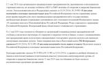
Workflows in a histology laboratory represent the orderly path that incoming material must follow to produce a histological report (Fig. 1). This path consists of a set of strictly mandatory and, in some cases, mandatory stages; operations at these stages are specific to the histological laboratory and discrete (intermittent). Not only is there no such thing as a “histology conveyor”, where on one side of an imaginary device one could place a container with material, and on the other side receive a response in one form or another (verbal, digital or some other), but also Many stages of histological examination are still not automated.
The histological material changes its shape twice as it passes through the stages of the study.
After the histological cutting step, representative sections travel throughout the laboratory in the form of histological blocks.
A histological block is a preparation embedded in a special paraffin medium, usually in the shape of an irregular cube (hence the name “block”). And after the microtomy stage - a process that involves cutting tissue for analysis with the required thickness of 2 to 5 microns - in the form of a preparation on a glass slide (the professional name for such a preparation is “glass”).
In this case, the received material can be divided into several blocks, and several glasses can be made from each block.
The drug ends its journey in a specialized archive, which is an important part of the laboratory. (Figure 1)
Rice. 1. Sequence of work processes in the histology laboratory
According to the regulations in force in our country, the patient’s drug (both the block and the glass) are subject to indefinite storage (the tissue in the paraffin block practically does not change its morphological, biological and chemical characteristics over time). The logical basis for this is clear - the histological conclusion confirms the need for surgical intervention, or the appointment of a specialized type of treatment, and it is not clear when exactly such confirmation may be needed. On the other hand, cancer patients, unfortunately, can return after primary treatment with relapses or complications, and in order to find out what is happening to the patient (relapse of a previously treated tumor, new tumor, metastasis), it is necessary to have a complete picture of the medical history, including previously collected material.
2.2 Risk factors associated with working conditions
Risk factors associated with working conditions include poor room lighting, low or high temperature indoors, poor air ventilation, high noise levels, stressful layout of workspaces and equipment, poor orientation in buildings, lack of personal protective equipment, ionizing radiation, electromagnetic fields and radiation, lack of laboratory security.
When assessing working conditions, injury safety assessment plays an important role. It is carried out on the basis of inspection of equipment (documentation for equipment), tools and devices, as well as the availability and completion of labor safety briefing logs and relevant entries in them for compliance with regulatory documents.
Among the risk factors, emotional and stress loads should also be assessed. Most health care facilities lack qualified personnel and overburden existing staff.
The laboratory should be illuminated with natural or artificial light, the level of which should be optimal for safe work. In addition, distracting and glare of light must be minimized.
Any equipment must be isolated from the general workspace if it emits excessive heat or cold. Personnel should be provided with personal protective clothing, including heat-protective gloves and appropriate clothing, to ensure comfort and personal safety.
The ambient temperature in the laboratory should be kept as comfortable as possible for laboratory workers.
Any equipment capable of emitting excess smoke, steam, heat, odors or toxicity should be placed under a suitable fume hood and isolated from the general work area. If this is difficult to accomplish, then it is necessary to carry out partial special re-equipment to create comfortable conditions for workers.
In case, as a result of certain processes self made If unpleasant or nauseating odors are formed, the use of mechanical or local natural ventilation is recommended.
Air movement and environmental humidity in the laboratory must meet the safety requirements of laboratory workers and be comfortable.
Air flow rates in the laboratory should prevent the dispersion of potentially infectious agents and toxic fumes and provide adequate ventilation.
The ventilation ducts of fume hoods should be separated from the general workspace to avoid dispersion or airborne transfer of infectious agents or odors to another part of the workspace.
There should be no unnecessary noise in the workplace. It is necessary to take into account the influence of individual elements of equipment on the overall noise level in the workplace when selecting and placing items. Appropriate measures must be taken to reduce and muffle the resulting noise generation.
Workspaces, laboratory activities, and equipment (eg, chairs, laboratory work stations, computer keyboards, and monitors), as well as vibrating and ultrasound equipment, should be located to reduce the risks of ergonomic distress-related disorders or accidents.
All laboratories that work with living biological agents must be equipped with design characteristics, taking into account compliance personal protection from moderate to high risk microorganisms. Laboratories that work with high-risk organisms should be equipped with design features that are more protective.
Laboratories must have emergency exits and signs at each entrance and exit. Signage at each location must combine internationally accepted hazard indicators (eg, biohazard, fire hazard, radioactivity) and other safety regulations.
The laboratory must have lockable doors. In emergency situations, locking the doors should not prevent egress. Access to the laboratory is limited to authorized personnel only. Locks may also be required on interior doors to prevent personnel from entering during processing of high-risk samples. When high-risk samples, cultures, chemicals or devices are stored, additional security measures are required, such as locked doors, locked refrigerators, restricted access of unauthorized persons to the laboratory, etc. Appropriate measures must be taken to prevent the threat of theft or damage to biological agents, samples, medications, chemicals or confidential information.
2.3 Biological risk factors associated with working in a histology laboratory
Working in a histology laboratory is associated with a number of hazards due to:
A) autopsy of people who died from various diseases, meaning infectious;
c) constant use in work of substances harmful to the body such as formaldehyde, chloroform, xylene, toluene, benzene, dioxane, mercury salts, aniline, etc.
d) use of flammable substances in work - alcohol, ether, etc.
The effect of formaldehyde on the human body.
Formaldehyde (from the Latin formoca "ant") is a colorless gas with a pungent odor, highly soluble in water, alcohols and polar solvents. Irritant, toxic. Formaldehyde is the first member of the homologous series of aliphatic aldehydes, formic acid aldehyde.
Application. An aqueous solution of formaldehyde (methanediol) stabilized with methanol - formaldehyde - causes denaturation of proteins, therefore in the leather industry it is used as a tanning agent and in the production of film for tanning gelatin. Formaldehyde is a good antiseptic due to its strong tanning effect. REMOVE This property of formalin is used in medicine: in the form of an antiseptic (Formidron, Formagel and similar preparations) and for the creation of anatomical and other preparations as a preservation of biological materials
One of the main sources of formaldehyde and urea in the production of urea-formaldehyde, melamine-urea-formaldehyde resins and for the treatment of urea against caking is an aqueous solution of formaldehyde (methanediol) stabilized with urea - KFK; for the production of plywood, chipboard is used in the woodworking and furniture industries, etc.
The main part of formaldehyde is used to produce thermoset polymers in the form of phenol-formaldehyde, urea-formaldehyde and melamine-formaldehyde resins. It is often used in the production process in industrial organic synthesis (pentaerythritol, trimethylolpropane, etc.). At temperatures below 9 during storage, the formaldehyde solution becomes cloudy and a white precipitate forms in the form of paraformaldehyde.
Formaldehyde is toxic. When exposed to the body, symptoms of chronic poisoning appear. A dose of 60-90 ml taken orally is lethal. Symptoms of foraldehyde poisoning: loss of strength, pallor, loss of consciousness, depression, breath holding, headache, and often night cramps.
In the case of acute inhalation poisoning, conjunctivitis and acute bronchitis appear, which subsequently develops to pulmonary edema. Dizziness, fear, unsteady gait, convulsions, symptoms increase gradually as the central nervous system is damaged. Poisoning through the mouth causes burns to the mucous membranes digestive tract. Swelling of the larynx and reflex cessation of breathing occurs.
There is pain in the throat, a burning sensation along the esophagus, pain in the throat, and if it gets into the stomach, vomiting of bloody masses and diarrhea occur. Hemorrhagic nephritis and anuria develop.
Those working with technical formaldehyde develop chronic poisoning, which is manifested by weight loss and dyspeptic symptoms. Damage to the central nervous system manifests itself in the form of persistent headache, poor sleep, mental agitation, tremors, ataxia, and visual disturbances. Organic diseases of the nervous system (thalamic syndrome) have been described; sweating disorder and temperature asymmetry occur. Cases of bronchial asthma are possible. Under the influence of formaldehyde vapors, say, among workers engaged in the manufacture of artificial resins or when direct contact with formaldehyde or its solutions, pronounced skin lesions in the form of dermatitis of the face, forearms and hands, fragility, and softening of the affected nails are observed, especially in the first days of work. The appearance of dermatitis and eczema is associated with the allergic nature of the exposure. After poisoning, there is an increased sensitivity to formaldehyde. There is scientific evidence indicating an adverse effect on specific functions of the female body.
Formaldehyde is included in the list of carcinogenic substances GN 1.1.725-98 in the section “probably carcinogenic to humans.” Its carcinogenic effect on animals has been fully proven. The connection between formaldehyde, used in the production of resins, plastics, paints, textiles, as a disinfectant and preservative has been proven (according to official data from the International Agency for Research on Cancer). There is an increased risk of developing cancer of the nasopharynx.
The effect of chloroform on the human body.
Chloroform (also known as trichloromethane, methyl trichlorimd, coldomn 20) is an organic chemical compound with the formula CHCl3. Under normal conditions, it is a colorless volatile liquid with an ethereal odor and a sweet taste. Practically insoluble in water. It forms solutions with water with a mass fraction of up to 0.23%. Chloroform is miscible with most organic solvents. Phosgene poisoning may occur when working with chloroform, which has been stored in a warm place for a long time.
Application. In the late 19th and early 20th centuries, chloroform was used as an anesthetic during surgical operations. Chloroform was first used as an anesthetic for surgical operations in England by physician Simpson (1848). In Russia, N. I. Pirogov was the first to use chloroform as a means for general anesthesia. In the future, chloroform was later replaced as an anesthesia by safer substances.
Chloroform is used to produce freon (freon-22). Chlorodifluoromethane. When obtained by the exchange reaction of chlorine atoms for fluorine as a treatment of chloroform with anhydrous hydrogen fluoride in the presence of antimony(V) chloride (according to the Swarts reaction). Chloroform is used as a solvent in industry to produce dyes and pesticides.
Chloroform contains deuterium (CDCl3). It is a solvent often used in nuclear magnetic resonance (NMR).
In the process of cleaning. First, chloroform combined with sulfuric acid is shaken, washed with water, dried over calcium chloride or magnesium sulfate, then distillation follows. Chloroform must be checked for purity by evaporation from filter paper. If chloroform is present, there should be no odor left. When a musty, pungent, irritating odor remains, this proves the presence of impurities of chlorine, hydrogen chloride or phosgene.
When exposed to the body, inhalation of chloroform has a detrimental effect on the functioning of the central nervous system. With a chloroform content of about 0.09% (900 ppm) in the air, dizziness, fatigue and headache can occur in a short time. Long-term toxic exposure to chloroform may contribute to the development of liver and kidney disease. An allergic reaction to chloroform entails an increase in body temperature (up to 40 °C) - the presence of such a reaction has been proven in approximately 10% of the population. Chloroform often causes vomiting - the incidence of postoperative vomiting is on average 75-80%.
In rats and mice that breathed air containing 0.003% chloroform (30 ppm), animal studies have proven the dependence of chloroform on the course of pregnancy and that miscarriages occurred. This has been observed in rats given oral chloroform. The effect on offspring has been proven: the next generations of rats and mice that inhaled chloroform had a higher percentage of birth defects than healthy individuals.
The effect of chloroform on offspring in humans is poorly understood. Possible death with prolonged exposure to the respiratory tract and mucous membranes of humans (2-10 minutes). Chloroform is suspected to be mutagenic and carcinogenic. Such properties appear when the permissible concentration of chloroform in the air is exceeded.
Once ingested, chloroform is eliminated fairly quickly through exhaled air: after 15-20 minutes. - 30-50% chloroform, within an hour - up to 90%. The remaining chloroform in the body is converted into carbon dioxide and hydrogen chloride as a result of biotransformation. The effect of xylene on the human body.
Xenobiotics (from the Greek oEnpt - alien and vYapt - life) are chemical substances alien to living organisms that are naturally not included in the biotic cycle. This is a conditional category for designation.
Because of economic activity human there is an increase in the concentration of xenobiotics in environment. Pesticides, some detergents(detergents), radionuclides, synthetic dyes, polyaromatic hydrocarbons, etc. - this is an incomplete list of xenobiotics. When released into the environment, drugs can cause allergic reactions, death of living organisms, changes the variability of hereditary traits, lowers immunity, disrupts the entire spectrum of metabolism, disrupts the course of processes in natural ecosystems, affecting the biosphere as a whole.
Examples of xenobiotics - xylene, styrene, toluene, acetone, benzene, gasoline vapor or hydrogen chloride - can be classified as xenobiotics at the moment when the maximum permissible concentration exceeds the norm. This accumulation in the environment in much higher concentrations can occur during industrial production.
The biological effect of xenobiotics, for example, with a slight inhalation of benzene vapor, immediate poisoning does not occur. For a long time, the procedure for working with benzene was not particularly controlled.
With large doses of benzene, nausea and dizziness occur, and in some cases, severe poisoning can lead to death. Euphoria is the first sign of benzene poisoning. Benzene vapors can penetrate the upper layers of the skin. Liquid benzene may cause skin irritation. With prolonged exposure to benzene in small quantities on the human body, the consequences become serious. A strong carcinogen can cause leukemia and anemia as a cause of chronic benzene poisoning.
Acute poisoning. At ultra-high concentrations, instant loss of consciousness and lightning death occur. Bluish coloration of the face, mucous membranes acquire a cherry-red hue. At lower concentrations - alcohol-like excitement, then drowsiness, malaise, general weakness, dizziness, nausea, vomiting, headache, accompanied by loss of consciousness. Unsustainable muscle twitching can develop into tonic spasms. The pupils are often dilated and do not respond to light. Increased breathing gives way to stopping. Body hypothermia decreases sharply. Tachycardia, rapid pulse, low filling. Blood pressure decreases. There may be cases of heart rhythm disturbances.
Long-term health disorders are observed after severe poisoning; sometimes they do not lead directly to death. There may be pleurisy, inflammation of the upper respiratory tract, eye diseases - cornea and retina, toxic liver damage, cardiac disorders, etc. A case is described a short time after acute poisoning with benzene vapors. Vasomotor neurosis occurs with swelling of the face and limbs, sensitivity disorders and convulsions; in such cases, death occurs a long time after poisoning.
In case of chronic poisoning, headaches, increased fatigue, shortness of breath, dizziness, weakness, nervousness occur, sleep suffers: drowsiness or insomnia, digestive disturbances: nausea, sometimes vomiting, loss of appetite, increased frequency of urination, menstruation, and persistent bleeding from the mucous membrane may occur. mouth, especially gums, and nose. Bleeding can last for a long time, hours or even days. Sometimes persistent bleeding occurs after tooth extraction. Small petechial hemorrhages in the skin are characteristic. Blood in stool, uterine bleeding, retinal hemorrhages. Bleeding itself and the accompanying hyperthermia (temperature up to 40° and above) leads to hospitalization. In such cases, the prognosis is always serious. Secondary infections are layered: there are known cases of gangrenous inflammation of the periosteum and necrosis of the jaw, there may be ulcerative inflammation of the gums, general sepsis with septic endometritis.
In severe poisoning, symptoms of damage to the nervous system occur. Variety of symptoms: Increased tendon reflexes, bilateral clonus, positive Babinski sign, deep sensory disorder, disorders with paresthesia, ataxia, paraplegia and motor disorders (signs of damage to the posterior columns of the spinal cord and pyramidal tracts).
blood changes are most typical. The number of red blood cells is usually sharply reduced. Hemoglobin levels drop. The color index in some cases is low, sometimes close to normal, and sometimes high (with blood thickening during bleeding). Anisocytosis and poikilocytosis and the appearance of nucleated red blood cells, an increase in the number of reticulocytes and the volume of red blood cells are noted. Accompanied by a sharp decrease in the number of leukocytes. Sometimes initially leukocytosis, quickly replaced by leukopenia, accelerated ESR. Changes in the blood do not develop simultaneously. Most often, the leukopoietic system is affected first, and thrombocytopenia occurs later. Erythroblasts are affected even later. As a result, a picture of severe poisoning occurs in the form of aplastic anemia.
Dry skin, cracks, itching are observed with frequent contact of hands with benzene. There may be redness (usually between the fingers), swelling, and millet-like blistering rashes. The maximum permissible concentration is 5 mg/m3.
As a rule, xenobiotics inhibit, to one degree or another, the nonspecific resistance of the organism. Humoral and cellular immune responses are affected. There are various hypersensitivity reactions (types 1-5, certain reactions of the immune system in this case increase). These reactions may be a manifestation of the immunotoxicity of xenobiotics, as well as suppression of the immune response. It happens when one of the components ensuring immune homeostasis increases while others are suppressed.
The effect of ethanol (alcohol) on the human body
Ethanol (ethyl alcohol, often colloquially simply “alcohol”) is a monohydric alcohol with the formula C 2H 5O H (empirical formula C2H6O), another option: CH3-CH2-OH, the second representative of the homologous series of monohydric alcohols, volatile under standard conditions, flammable, colorless transparent liquid. Ethyl alcohol is also used as a fuel, as a solvent, as a filler in alcohol thermometers, and as a disinfectant (or as a component thereof).
Application.
In medicine, ethyl alcohol is primarily used as a solvent, extractant and antiseptic, as a disinfectant and drying agent, externally; the drying and tanning properties of 96% ethyl alcohol are used to treat the surgical field or in some techniques for treating the surgeon’s hands;
solvent for medicines, for the preparation of tinctures, extracts from plant materials, etc.;
preservative for tinctures and extracts (minimum concentration 18%);
defoamer when supplying oxygen, artificial ventilation;
in warm compresses;
for physical cooling during fever (for rubbing);
component of general anesthesia in situations of drug shortage;
as an antifoam for pulmonary edema in the form of inhalation of a 33% solution;
ethanol is an antidote for poisoning with certain toxic alcohols such as methanol and ethylene glycol. Its action is due to the fact that the enzyme alcohol dehydrogenase, in the presence of several substrates (for example, methanol and ethanol), carries out only competitive oxidation, due to which, after timely (almost immediate, following methanol/ethylene glycol) intake of ethanol, the current concentration of toxic metabolites decreases (for methanol - - formaldehyde and formic acid, for ethylene glycol - oxalic acid)
...Characteristics of microbial air pollution in dental offices. Comprehensive assessment working conditions for dentists. Psychophysical indicators of the body medical workers at the beginning and end of the working day. Assessment of health risk factors for doctors.
abstract, added 12/22/2015
Health care system today. Training and instruction of workers on labor protection. The main harmful production factors of working conditions. Chemical hazards used in medical practice, the effects of drugs, physical factors.
abstract, added 03/27/2010
Characteristics of the X-ray room of the regional tuberculosis hospital, institution IK-4. Analysis of the types of studies carried out in the X-ray room. The procedure for preliminary assessment of layer-by-layer images of the lungs. Sanitary and anti-epidemic measures.
abstract, added 12/16/2013
Labor theory about the origin of man. The impact of internal and external factors on skeletal development. The relationship between the social and the biological in the structure of bones. The influence of sports on changes in the composition, processes of growth and ossification of bone.
presentation, added 05/21/2014
Concepts of labor physiology. Physiological characteristics of mental work. Redistribution reactions of cerebral blood flow. The relationship between mental and physical labor. Hygienic criteria for assessing labor depending on the severity of the labor process.
abstract, added 07/02/2013
Study of the main symptoms and clinical course of chronic viral hepatitis. Study of factors determining disease progression and the effectiveness of antiviral therapy. Analysis of the incidence of chronic hepatitis in the Primorsky Territory.
course work, added 10/06/2016
Unfavorable labor factors of various groups of medical workers. Conditions and features of occupational hygiene of individual specialties. Hygienic assessment of the work of medical workers using ultrasound equipment. The degree of severity and intensity of work.
presentation, added 11/23/2014
The objectives of therapeutic and preventive nutrition are the use of specially formulated food rations and diets for therapeutic purposes. Prevention of adverse effects production factors. Working conditions according to the degree of harmfulness and danger.
Staining of preparations
Basic paints Karazzi and hematoxylin Mayer.
Acid paints -
Simple - one paint is used
Double - two colors are used
Complex - three colors are used
histochemical coloring:
On the slime
For amyloid - Congo mouth is red.
Fat staining –
Identification of iron compounds.
(According to Perls)
(According to Tirman)
(Chic reaction)
Staining of bacteria in sections.
According to Gram.
Orders, resolutions, instructions regulating work in PJSC
1 Federal Law No. 8-F3 dated January 12, 1996 (as amended on November 25, 2009) “On consumption and funeral business” (adopted by the State Duma of the Federal Assembly of the Russian Federation on December 8, 1995)
2 Order of the Ministry of Health and Medical Industry of Russia dated April 29, 1994 No. 82 “On conducting pathological autopsies”
3 List of accounting and reporting medical documentation of PJSC approved by order of the Ministry of Health Russian Federation dated October 4, 1980 No. 1030
4 Instructions for organizing work and observing the anti-epidemic regime of PJSC and branches of the Forensic Medical Examination Bureau in cases of suspicion or detection of particularly dangerous infections(approved by the Deputy Ministry of Health of the USSR on December 12, 1978)
5 Rules for the arrangement and operation of the premises of pathological departments of morgues (pathological and forensic laboratories), treatment and preventive and forensic medical institutions, institutes and educational institutions (to SNiP 02.08.02-89)
6 Works and services in the specialty of pathological anatomy in the nomenclature of works and services for the provision of medical care (Order of the Ministry of Health of the Russian Federation dated July 26, 2002 No. 238)
REPORT on the work done by a medical assistant-laboratory assistant-histologist
Pathoanatomical Department of Federal State Healthcare Institution KB-122 named after. Sokolova L.G.
FMBA of Russia Neli Alexandrovna Platonova.
After graduating from the 6th Leningrad Medical School in 1972 with a specialty nurse, was assigned to work in 81 clinics in the city of Leningrad. Since October 1982 she worked at KDL TsMSCh-122. Since 1984, she worked in the urgent analysis laboratory as a laboratory assistant on duty. Since May 1996 I have been working in the pathology department as a medical assistant-laboratory assistant-histologist, I have a certificate as a laboratory assistant-histologist and a higher qualification category medical assistant-laboratory assistant-histologist.
Responsibilities of a medical assistant histologist:
Receive biopsied and surgical material delivered for research from clinical departments and the operating room.
Check the markings, computer number, code, compliance of the delivered material with the records in the accompanying direction.
Preliminarily wash the material with running water to remove formaldehyde.
Participate in cutting material.
Mark the cut pieces, record their quantity, methods of subsequent histological processing.
Fill out the referral form on the computer and a macro description of the material as dictated by the doctor.
Carry out wiring, paraffin embedding, histological processing, production of high-quality microslides and examined surgical and biopsy material.
Participate together with the doctor in cutting out fixed sectional material.
Pour the cut pieces into paraffin and other media.
Stain sections and prepare microslides.
Perform urgent production of histological microslides on a freezing microtome.
Ensure proper sanitary and hygienic conditions in the laboratory.
Maintain order and cleanliness in the workplace.
Improve your qualifications in a timely manner.
The pathology department is a structural subdivision of KB-122 named after. Sokolova L.G FMBA of Russia.
The pathology department includes laboratories:
Histological laboratory- examines surgical, biopsy, sectional material coming from departments of KB-122
In 2016, 9192 patients were examined, 82228 examinations of surgical and biopsy material were performed, of which:
Total: I completed:
Operating material 47862 kus. / 6258 kus.
FGS biopsies 10345 kus. / 1515 kus.
Endometrial scrapings 17802 kus / 3103 kus.
Biopsies 6219 kus / 703 kus.
54 urgent biopsies performed
77 autopsies performed
Cytology laboratory-
performs cytological and cytochemical studies of material coming from departments of KB-122
In 2016, 12,274 patients were examined, 24,048 cytological studies were performed:
Laboratory of immunohistochemical research methods -
performs and determines with high accuracy the nature of tumor processes, differentiates the degree of malignancy and sensitivity of tumor cells to hormonal and chemotherapy using special markers. The laboratory was organized in 2009.
In 2016, 601 patients were examined, 1838 immunohistochemical studies of surgical and biopsy material were performed.
Performed according to job descriptions next job:
Excision of surgical and biopsy material
The material is delivered to the laboratory in fixed form in a 15% formalin solution with a direction. For immunohistochemical studies, the material is delivered to the laboratory immediately after surgery not fixed.
1. All material is sorted into surgical, biopsy, FGS biopsies and endometrial scrapings.
2. Registered in an alphabetical journal and entered into the computer. Each individual case is given its own number, which makes it possible to record not only the number of pieces, but also the number of patients per year.
3. The doctor dictates a description of the surgical material (macropreparation - macrodescription). Then the material is put into wiring.
4. The wiring takes place in an Automatic tissue processor with software control for wiring the histological material.
5. Surgical and biopsy material is embedded in the LEICA 1150 paraffin embedding station.
6. The material is then poured and cut using a microtome. After cutting, the material is placed in a thermostat at t-42* overnight, and then the preparations are stained.
7. Urgent biopsy material is cut on a LEICA RM 2125 Cryostat with further staining. The time for urgent research does not exceed 30 minutes. In case of an urgent biopsy, the doctor gives a preliminary opinion. The final answer is given based on the material filled in.
Staining of preparations
All paints used in histology are divided into nuclear or (basic) and diffuse (acidic). For the vast majority of diagnostic studies, the following are sufficient simple ways coloring like Hematoxylin-eosin and Van Gieson.
Basic paints. Of the nuclear (basic) paints, paints made from hematoxylin are most widely used. Our laboratory uses hematoxylin Karazzi and hematoxylin Mayer.
Acid paints - Eosin, acid fuchsin, and picric acid are constantly used. The last two paints are used in a special mixture with each other, called picrofuchsin or Van Gieson mixture.
In histological practice, depending on the number of dyes used, the following types of dyes are distinguished:
Simple - one paint is used
Double - two colors are used
Complex - three colors are used
In all these colors, the main role belongs to the nuclear (basic) paint, which is used either alone or in combination with acidic ones (one or more). In histological practice, hematoxylin is used in combination with eosin (double) and hematoxylin with picrofuchsin (triple) - Van Gieson.
In some cases they are used histochemical coloring:
On the slime– has great diagnostic value in some pathological processes - tumors, callogenosis.
Alcian blue, mucicarmine.
For amyloid - To detect amyloid, there are a number of special methods, of which the most common are Congo mouth is red.
Fat staining – Sudan III, Sulphate Nile blue.
When staining for fat, all sections must be after freezing.
Identification of iron compounds.
The material to be tested for iron must be fresh.
There are two reactions to detect iron:
Reaction to Prussian blue(According to Perls)
Reaction to turbulene blue(According to Tirman)
Iron salts in both cases give blue compounds.
Coloring of pathogenic fungi and neutral mucus.
(Chic reaction)
Staining mushrooms with fuchsulfuric acid - Schiff reaction.
Staining of bacteria in sections.
Stained with Lefler's methylene blue.
According to Gram.
We bring to your attention a typical example of a laboratory assistant job description, sample 2019/2020. Job description of a laboratory assistant should include the following sections: general position, job responsibilities of a laboratory assistant, rights of a laboratory assistant, responsibility of a laboratory assistant.
The laboratory assistant's job description should reflect the following points:
Job responsibilities of a laboratory assistant
1) Job responsibilities. Performs laboratory analyses, tests, measurements and other types of work during research and development. Participates in the collection and processing of materials during the research process in accordance with the approved work program. Monitors the good condition of laboratory equipment and adjusts it. Prepares equipment (instruments, equipment) for experiments, carries out its checks and simple adjustments in accordance with developed instructions and other technical documentation. Participates in the implementation of experiments, carries out the necessary preparatory and auxiliary operations, conducts observations, takes instrument readings, and maintains work logs. Provides department employees with the equipment, materials, reagents, etc. necessary for work. Processes, systematizes and formalizes in accordance with methodological documents results of analyses, tests, measurements, keeps records of them. Selects data from literary sources, abstracts and information publications, normative and technical documentation in accordance with the established task. Performs various computational and graphical work related to ongoing research and experiments. Participates in the preparation and execution of technical documentation for the work performed.
The laboratory technician should know
2) When performing his/her duties, a laboratory technician must know: guidelines, normative and reference materials related to the topic of work; methods of analysis, testing and other types of research; current standards and technical specifications on the technical documentation being developed, the procedure for its preparation; laboratory equipment, control and measuring equipment and rules of its operation; methods and means of performing technical calculations, computational and graphic work; basics of economics, organization of labor and production, operating rules computer technology; basics of labor legislation; internal labor regulations; rules and regulations of labor protection.
Laboratory assistant qualification requirements
3) Qualification requirements. Average vocational education without presenting requirements for work experience or initial vocational education and work experience in the specialty of at least 2 years.
1. General provisions
1. The laboratory assistant belongs to the category of specialists.
2. A person who has a secondary vocational education without requirements for work experience or primary vocational education and work experience in the specialty of at least 2 years is accepted as a laboratory assistant.
3. A laboratory assistant is hired and dismissed _____ (director, manager) organization represented by _____ (position).
4. The laboratory assistant must know:
- guidelines, normative and reference materials related to the topic of work;
- methods of analysis, testing and other types of research;
- current standards and technical specifications for the technical documentation being developed, the procedure for its preparation;
- laboratory equipment, control and measuring equipment and rules of its operation;
- methods and means of performing technical calculations, computational and graphic work;
- basics of economics, organization of labor and production, rules of operation of computer equipment;
- basics of labor legislation;
- internal labor regulations;
- rules and regulations of labor protection.
5. In his activities, the laboratory assistant is guided by:
- legislation of the Russian Federation,
- Charter (regulations) of the organization,
- orders and instructions _____ (general director, director, manager) organizations,
- this job description,
- Internal labor regulations of the organization,
6. The laboratory assistant reports directly to: _____ (position).
7. During the absence of a laboratory assistant (business trip, vacation, illness, etc.), his duties are performed by a person appointed ______ (position) of the organization in the prescribed manner, who acquires the corresponding rights, duties and is responsible for the performance of the duties assigned to him.
2. Job responsibilities of a laboratory assistant
Laboratory assistant:
1. Performs laboratory analyses, tests, measurements and other types of work during research and development.
2. Participates in the collection and processing of materials during the research process in accordance with the approved work program.
3. Monitors the good condition of laboratory equipment and adjusts it.
4. Prepares equipment (instruments, equipment) for experiments, carries out its checks and simple adjustments in accordance with developed instructions and other technical documentation.
5. Participates in the implementation of experiments, carries out the necessary preparatory and auxiliary operations, conducts observations, takes instrument readings, and maintains work logs.
6. Provides department employees with the equipment, materials, reagents, etc. necessary for work.
7. Processes, systematizes and draws up the results of analyzes, tests, measurements in accordance with methodological documents, and keeps records of them.
8. Selects data from literary sources, abstracts and information publications, normative and technical documentation in accordance with the established task.
9. Performs various computational and graphical work related to ongoing research and experiments.
10. Participates in the preparation and execution of technical documentation for the work performed.
3. Rights of a laboratory assistant
The laboratory assistant has the right:
1. Submit proposals for management’s consideration:
- to improve work related to those provided for in this instructions and duties,
- on the encouragement of distinguished employees subordinate to him,
- on bringing to material and disciplinary liability workers who violated production and labor discipline.
2. Request from structural divisions and employees of the organization information necessary for him to perform his job duties.
3. Get acquainted with the documents defining his rights and responsibilities for his position, criteria for assessing the quality of performance of official duties.
4. Get acquainted with the draft decisions of the organization’s management relating to its activities.
5. Require the management of the organization to provide assistance, including ensuring organizational and technical conditions and execution of the established documents necessary for the performance of official duties.
6. Other rights established by the current labor legislation.
4. Responsibility of the laboratory assistant
The laboratory assistant is responsible in the following cases:
1. For improper performance or failure to fulfill one’s job duties provided for in this job description - within the limits established by the labor legislation of the Russian Federation.
2. For offenses committed in the course of their activities - within the limits established by the current administrative, criminal and civil legislation of the Russian Federation.
3. For causing material damage to the organization - within the limits established by the current labor and civil legislation of the Russian Federation.
Job description for laboratory assistant - sample 2019/2020. Job responsibilities of a laboratory assistant, rights of a laboratory assistant, responsibility of a laboratory assistant.